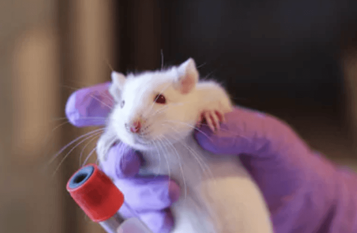
Tumor animal models can be divided into three categories: spontaneous tumor animal models, induced tumor animal model and animal models of transplantable tumors. The tumor-bearing mouse models that we can provide are mainly divided into two types. One is to inoculate human or mouse-derived cell lines into immunodeficient mice, called CDX models, and the other is to tumours derived from patients. Tissue pieces are inoculated into immunodeficient mice and are called PDX models.
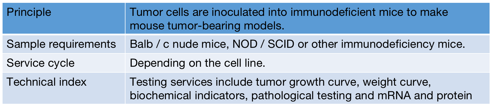
Tumor cell tumor bearing model Sample requirements
Tumor cell tumor bearing model Experimental data
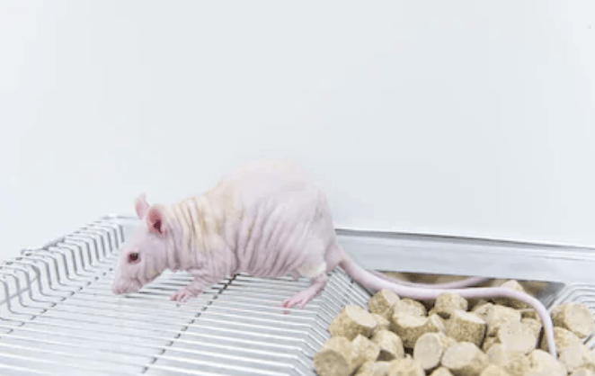
CDX tumor model in vivo
Luciferase-labeled tumor cell lines can be used to construct a variety of subcutaneous, in situ or metastatic CDX tumor models. The Luciferase reporter gene system uses luciferin as a substrate to detect the activity of firefly luciferase. The advantage is that it can be repeatedly measured while the mouse is alive, and the fluorescence changes at different time points can be analyzed. To trace tumor cell proliferation and metastasis.
Subcutaneous tumor formation model of nude mouse HepG2 cells
Transplanted liver cancer models commonly used in the laboratory can be divided into orthotopic and ectopic according to the location of the transplanted tumor. Heterotypes often form tumors by subcutaneous injection of liver cancer cells in experimental animals. The subcutaneous tumor formation experiment is simple in operation, high in success rate, and easy to measure tumor volume change, which is convenient for local intervention.Improved bioinformatics tools assist in the selection of new antigens by predicting the presentation of mutant peptides for recognition by the immune system.
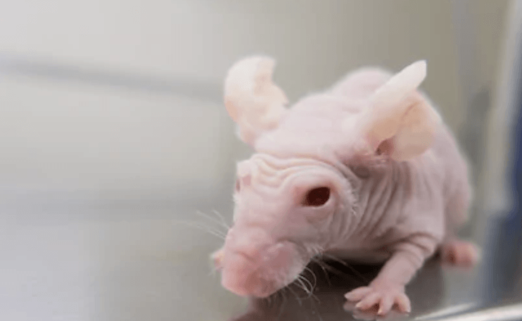
Hepatoma patient-derived xenograft model
Advantage:
PDX preserves the diversity of genotypes and phenotypes of the patient's tumor tissue, which truly reflects the characteristics of the original tumor;
PDX preserves tumor stromal cells and preserves tumor microenvironment;
Compared with tumor cell lines, the findings of PDX can more accurately reflect the tumorigenesis and development mechanism of patients.
Applied to:
Screening of anticancer drugs and development of biomarkers; collaborative clinical trials of oncology drugs; precision medicine; tumor mechanism.
Available PDX Services:
Self-produced immune-deficient M-NSGTM mice provide PDX transplant recipients; mature subcutaneous and organ inoculation techniques ensure F0 PDX construction and passage expansion; pathological and molecular biology techniques ensure PDX identification; cell culture techniques ensure long-term PDX Cryopreservation
Our advantage
Mature and reliable Search for Tumor Animal Models Solutions platform: rich project experience, participate in major projects, and publish high-level cooperation results.
The analysis contents and methods based on high-quality data analysis, pre-research and verification are indeed mature and feasible.
Creative Proteomics leads a rich team that uses the most rigorous experimental solutions and internationally recognized experimental techniques for Tumor Animal Models to obtain your customized information. Use the most accurate experimental results to help you better carry out tumor-related scientific research.
* For Research Use Only. Not for use in the treatment or diagnosis of disease.
Related Services: