Metastasis is the cumulative result of many changes in tumor cells and their microenvironment, which enables cells to migrate and invade healthy host tissues. As proliferative tumor cells try to escape from the primary tumor site, local cell adhesion and surrounding tissue invasion must occur. Before penetrating the vascular endothelium and entering the bloodstream, cancer cells must invade local tissue by degrading ECM protein components and eventually cross the basement membrane. Once cycled, these cells can form metastatic colonies in secondary locations.
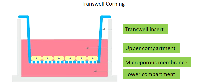
Transwell is a kind of permeable cup-shaped device. After being put into the culture plate, it is divided into two chambers: the Transwell chamber is called the upper chamber, which is filled with the upper culture medium; the culture plate is called the lower chamber, which is filled with the lower culture medium. The culture medium of the upper and lower layers was separated by polycarbonate membrane. Tumor cells are grown in the upper chamber, and because of the permeability of polycarbonate membrane, the effects of components in the lower culture medium on the growth and movement of tumor cells can be studied.We have high-quality researchers and experienced team to provide you with personalized tumor Transwell experimental solutions, with the most accurate experimental results to help you better carry out cancer-related scientific research.
Scope of application
1. Tumor migration experiment: to study the migration ability of tumor cells or the migration ability of tumor cells under specific conditions;
2. Tumor cell chemotaxis experiment: to study the effect of tumor cell secretory product or a certain factor / protein on the migration of another tumor cell;
3. Tumor invasion experiment: to study the invasive ability of tumor cells or the invasive ability of tumor cells under specific conditions.

(1) All tumor cell culture reagents and Transwell chamber were incubated at 37℃.
(2) The tumor cells to be tested were cultured to logarithmic growth phase, digested cells, suspended tumor cells in serum-free medium;
(3) 600-800μl medium containing 10% serum was added in the lower chamber, and 100-150 μl cell suspension was added in the upper chamber and cultured for 24 hours;
(4) Take out chamber, drain the liquid, move it to 800μl methanol hole and fix it for 30 minutes;
(5) Take out chamber, and dry the fixed solution, move it to the hole of 800μl Giemsa dye solution and dye it for 15 minutes;
(6) Rinse with clean water, remove chamber, to absorb the fluid from the upper chamber, and wipe off the cells on the surface of the membrane at the bottom of the upper chamber;
(7) Remove the film with tweezers and seal the film after drying;
(8) Nine random visual fields were counted under microscope and the results were counted.
Provided by the customer
1.Experimental tumor cells in good growth condition and cell culture conditions (dry ice is needed for cryopreserved cells.
To transport or culture tumor cell samples are sent in a culture bottle full of culture medium, and the bottle mouth is sealed.
2.It is necessary to accurately inform the contents of the experimental design, such as sample concentration, etc.
We provide
1.A full set of detailed experimental reports, including experimental flow, material methods, experimental results, etc.
2.The original data and pictures were detected by Transwell.
Service cycle
Chemotaxis (no Matrigel), for 10 working days;
Invasion (use Matrigel), for 10 working days.

Creative Proteomics leads a rich team that uses the most rigorous experimental solutions and internationally recognized transwell experimental techniques to obtain your customized information. Use the most accurate experimental results to help you better carry out tumor-related scientific research.
Reference
1. Song Y. et al. (2011). "Effects of RNA interference targeting four different genes on the growth and proliferation of nasopharyngeal carcinoma CNE-2Z cells." Cancer Cell, 18(4):297-304..
* For Research Use Only. Not for use in the treatment or diagnosis of disease.
Related Services: